Why Get Wellness Photos (Retinal Photo and OCT)?
It’s finally exam day and you’re so excited to have your eyes checked. Ok, maybe not, but at least hooray for new glasses! And then the tech asks if you want to have wellness photos of your retina (record scratch). Hmm. You’re not sure. Is that something the doctor wants? Is it something I need? What even is a retina? We’ve put together this guide to help you understand what this is and how it could enhance your exam depending on your specific needs. Read on to learn about how those with diabetes, hypertension, macular degeneration, certain cancers, glaucoma, and history of systemic medications like Plaquenil, Accutane, Tamoxifen, and Viagra/Cialis may want to seriously consider getting wellness imaging.
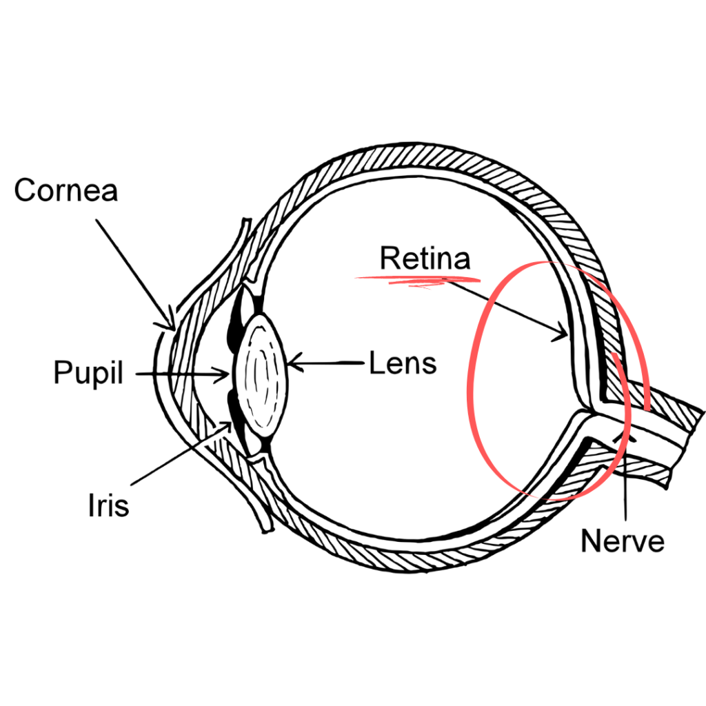
The retina is a tissue at the back of the eye made up of millions of cells that convert light waves to an electrical signal for the brain. The brain then processes this signal into what we perceive as vision. Any disruption to this tissue or the pathway will disrupt vision. Although, many disruptions to the retina do not affect vision at all. Thankfully, wellness imaging allows us to evaluate your eye health more precisely.
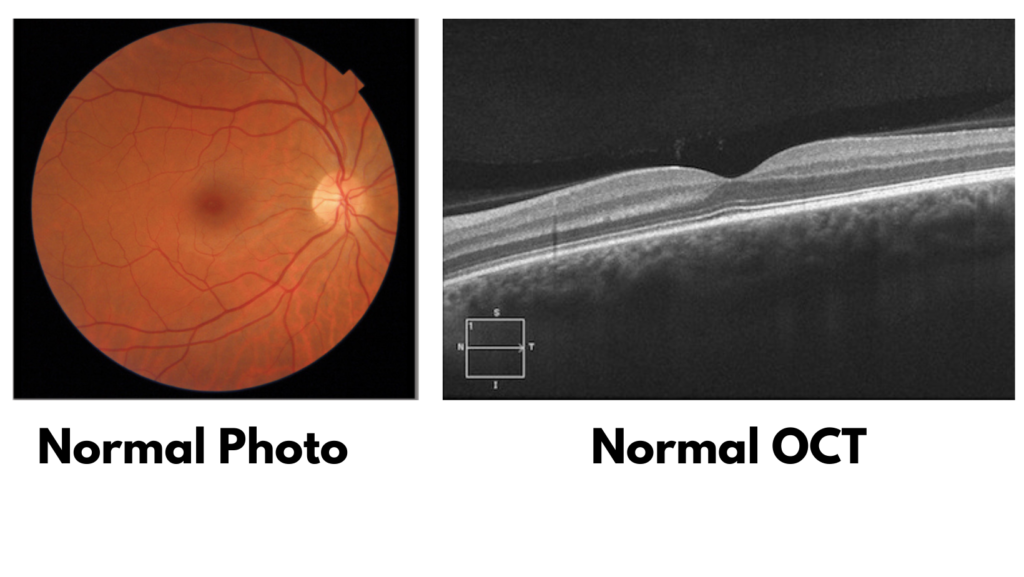
Retinal Photo and Retinal OCT
Wellness photos at The Eye Shop is actually more than just a simple picture of the retina. This camera is also equipped with Optical Coherence Tomography (OCT). That’s a lot of words that basically mean “ultrasound, but with light”. It gives your doctor two different views so they can more thoroughly inspect the health of your eyes.
It’s critical to point out that in each of the conditions discussed, it is possible – common even – to feel healthy and see 20/20 and have these complications. Bad vision and ocular complications are always great reasons to present for an eye exam, but do not assume 20/20 vision and normal feeling eyes is the same as “healthy eyes”.
Normal Eyes / Baseline Images
Surely not everyone has an explicit medical need for imaging. However, it is always beneficial to have a baseline image to refer to in case your eye health ever changes. Having a reference point from when the eye is healthy can be invaluable information to refer to if it is no longer healthy. For this reason, we recommend having at least one baseline image with us on file.
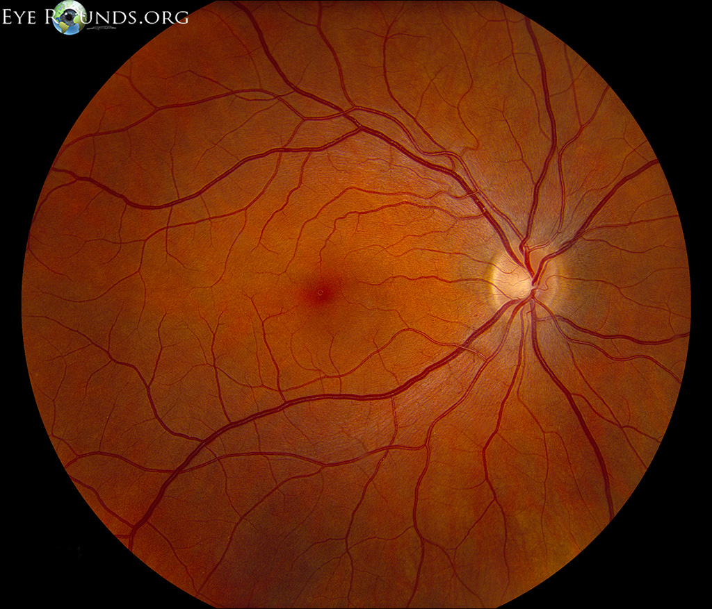
As we discuss imaging, please use these images below as a reference for what is considered normal. In the photo to the left, we like to refer to this as the Google Satellite image. We’ve got a nice birds-eye view of the back of your eye. The yellow circle to the right is your optic nerve head where 1.2 million nerve fibers leave your eye to go to the brain. The darker circle in the middle of the photo is your macula, the area that is responsible for processing your central vision.
The image to the right is an OCT of the eye and we refer to this as the Google Street View. We’re now *in* the layers of the tissue with a cross-sectional view. To orient, The black at the top of the image is the inside of your eyeball. The layered gray with the divot is your retina, with the divot being where your central vision is processed. The hazy, sponge-like tissue under that white line is the rest of the back of your eye. It may be helpful to refer back to the drawing of the retina featured above (the area circled in red). That’s what we’re looking at with an OCT.
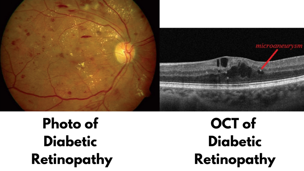
Diabetes
Ideally, all diabetic patients would get imaging at every visit. Diabetes is notoriously sneaky and can “quietly” have negative effects. Patients can have significant diabetic retinopathy even if they’re still seeing 20/20, which is why an annual exam is always necessary. Diabetic retinopathy is the leading cause of preventable blindness in working age adults and can be prevented with early detection. We firmly believe imaging for patients with diabetes is an essential part in managing your diabetic care and preserving your vision.
Macular Degeneration
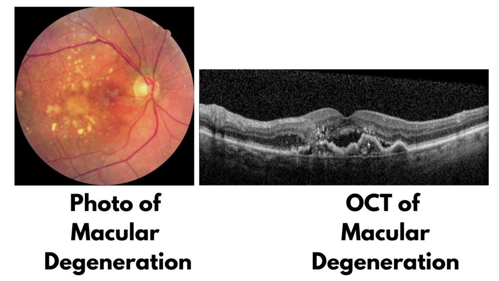
Patients regularly voice concerns with us over macular degeneration. Maybe they’ve seen a friend struggle with the condition or they know it runs in the family. Or maybe they have a diagnosis of macular degeneration and are working hard to keep it from the devastating “wet” form (leakage of blood or plasma). A photo with OCT is critical in these scenarios to give the doctor ample information to correctly diagnose and manage your case.
Freckles (choroidal nevus) / Melanoma / History of Certain Cancers
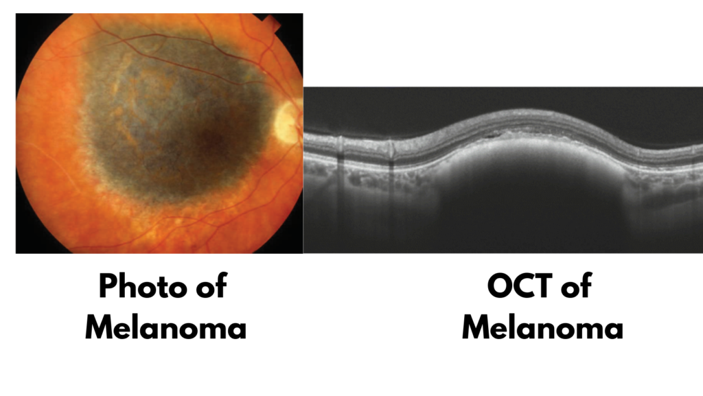
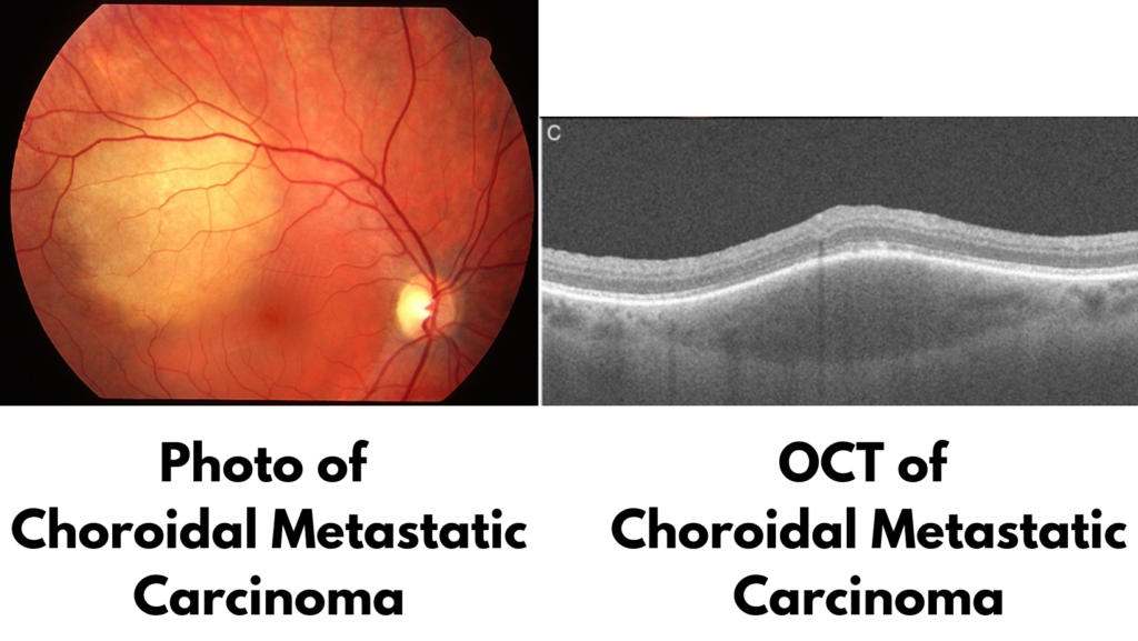
Something we hear a lot is “The doctor where I used to live said there was a freckle or a dot in the back of my eye and to get it checked every year”. Usually, this is in reference to a choroidal nevus and like any freckle or mole, it has the risk of converting to ocular melanoma, a devastating cancer of the eye. Additionally, any patient with a history of cancer may have secondary tumors in the eye (tumors that spread to the eye from another location in the body). Specifically, men with a history of lung cancer and women with a history of breast cancer are at risk for choroidal metastatic carcinoma. Whether it’s a previous freckle you’ve been told about or you’ve got a history of cancer, imaging is definitely recommended.
High Blood Pressure (Hypertensive Retinopathy)
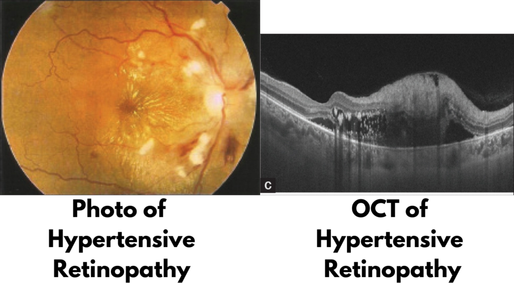
Nearly 80 million people in the US have high blood pressure (hypertension) and of those, one study shows barely over half are appropriately treated. Known as the “silent killer” due to the condition having almost no symptoms, many patients are in physical dire straits but feel totally normal. Risks include cancer, heart disease, and stroke. If well-controlled, the patient’s retinal health may look almost completely normal. However, poor control of hypertension can have devastating effects on the eyes, as well as systemic health.
Epiretinal Membrane
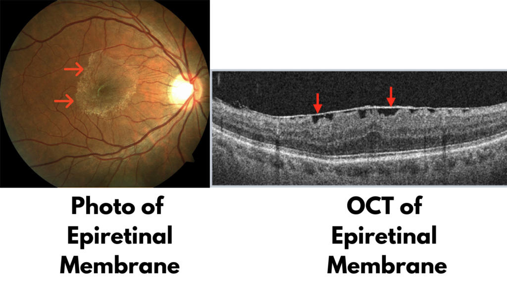
Epiretinal membrane is the condition so nice they named it thrice. It’s also known as “macular pucker” and “cellophane maculopathy”. Of all those options, we like the latter best because it really describes what the condition looks like AND does. It looks like a piece of cellophane on the retina and bunches up like cellophane on a countertop. It’s an extra layer that grows on top of the retina and disrupts the very specific orientation of light sensing cells in the retina, resulting in distorted vision. An OCT image is critical in the follow-up care and surgical consideration for this condition.
Issues with Medication: Plaquenil / Accutane / Tamoxifen / Viagra & Cialis
Patients are sometimes surprised to hear us ask about their systemic medications when they come in for an eye exam. However, many of the medications you take for non-ocular issues can have ocular impacts. And again, vision is not a great predictor for some of these issues. This is nowhere near an exhaustive list, but some common examples are:
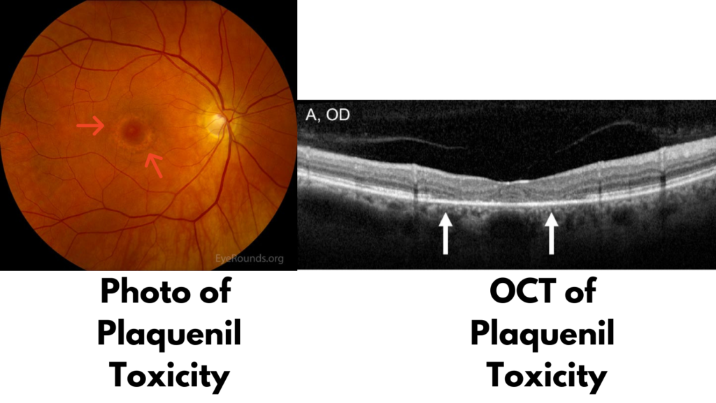
- Plaquenil (Hydroxychloroquine): This medication is commonly used for autoimmune disorders and is typically prescribed by a rheumatologist. That rheumatologist is also probably pretty specific about getting specific eye testing on a regular basis. And for good reason; this medication has widely known retinal toxicity that can affect your mid-peripheral vision. Patients may see 20/20, but find it impossible to do simple tasks that use mid-peripheral vision, like reading. See image below.
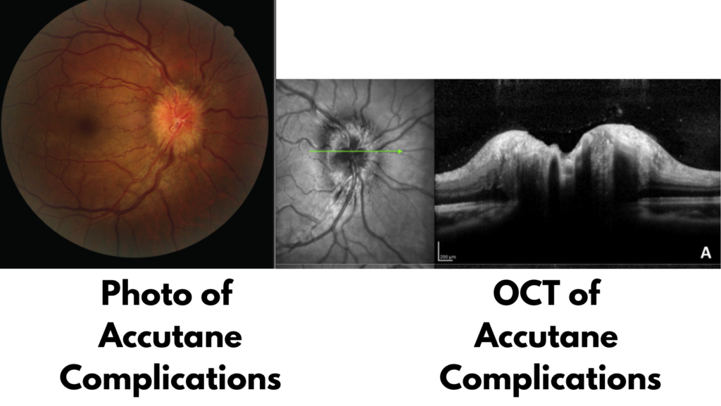
- Accutane (Isotretinoin): This medication is used for treating acne. The PCP or dermatologist that prescribes this medication may also require regular eye exams because it has a long list of ocular side effects. From tunnel vision, color defects, night vision issues, and more. But one of the issues seen with this medication is swelling to the optic nerve head. See image below.
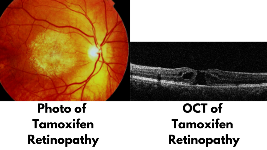
- Tamoxifen: This medication is used for treating breast cancer and also has a varied list of ocular side effects. The ones we’d like to capture on image and OCT are refractile deposits in the retina and macular cysts. See image below.
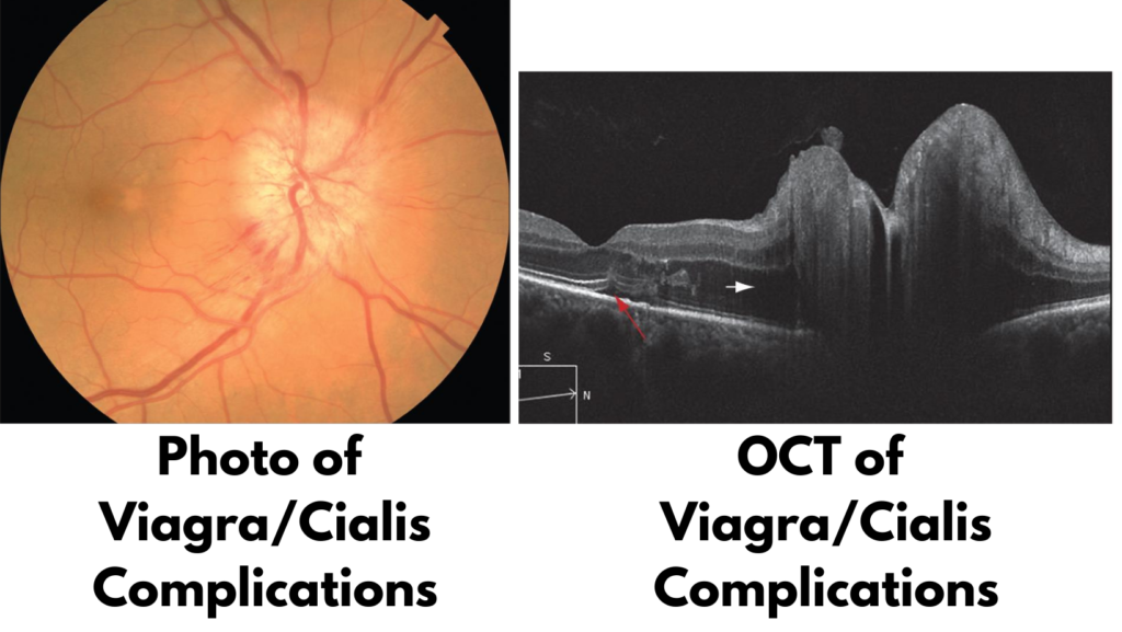
- Viagra (Sildenafil) & Cialis (Tadalafil): Among the ocular side effects of these erectile dysfunction medications is risk for developing Non-Arteritic Ischemic Optic Neuropathy (NAION), a blinding condition. The incidence is low and may be dependent on other factors, but due to the severity of outcome for those that suffer with it, caution is recommended. See image below.
Optic Nerve Issues / Suspicion of Glaucoma
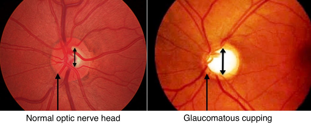
Has an eye doctor every talked to you about your optic nerves? Many patients tell us, “My old eye doctor was watching my nerves” or “I’ve been told one of my nerves is bigger but they didn’t seem concerned”. Usually this means your nerves had an appearance that made you a glaucoma suspect, but did not warrant a full glaucoma diagnosis. Other times it means there was something else unique about your nerves, like optic disc coloboma, multiple sclerosis, myelinated nerve fiber layer, trauma, or countless others. Either way, imaging is recommended. And naturally, all glaucoma diagnosed patients at The Eye Shop will get imaging to aid in the management of their condition.
Other Considerations
It would be impossible to cover every single condition when thinking about getting retinal photos done. Suffice it to say that there isn’t a bad reason to get wellness photos. Some patients are eager to get photos after they learn a family member has an eye condition like retinitis pigmentosa, histoplasmosis, strokes of the eye, and more. Sometimes they’re just curious to see what the inside of their eye looks like. Some patients just like the added peace of mind that comes with a more in-depth view of their eyes. As always, you’re definitely welcome to discuss it with your doctor when you come in for your exam. We are happy to explain why we would like the photos and, naturally, will review your photos with you so you can see what we see and learn more about what it is we want to document.
Cost
If your visit with us is under your vision insurance or you’re a cash pay patient, wellness photos are $59. If your visit is through an in-network medical plan and you are seeing us for a medical concern that requires imaging, we will bill your insurance.
For more information or to schedule your appointment, call us today at 480-656-7739.

Leave a Reply Cancel reply
follow us
schedule an appointment
website designed by katie & Co. Design
come visit us AT EITHER LOCATION!
The Eye Shop OCOTILLO
21321 E. Ocotillo #105,
Queen Creek, AZ 85142
THE EYE SHOP POWER
18550 E. RITTENHOUSE #100,
Queen Creek, AZ 85142
How much do you charge for a wellness photo?
Great question! As of this writing (September 2023), we charge $59 for wellness photos.
I so appreciate all the above information and particularly that it was sent to me prior to my exam. This gives me time to absorb the information and then happily/gratefully pay the fee for the photos. Looking forward to my exam.
I so appreciate all the above information and particularly that it was sent to me prior to my exam. This gives me time to absorb the information and then happily/gratefully pay the fee for the photos. Looking forward to my exam.